Examination and imaging
Diagnosis begins with imaging.
The part visible in the mouth is the crown of the tooth. The source of pain, whether a tooth can be saved and whether an implant is appropriate are determined by assessing the root, the root canal and the surrounding bone on a radiograph. As you scroll down, the film lifts and what is visible in the mouth remains.
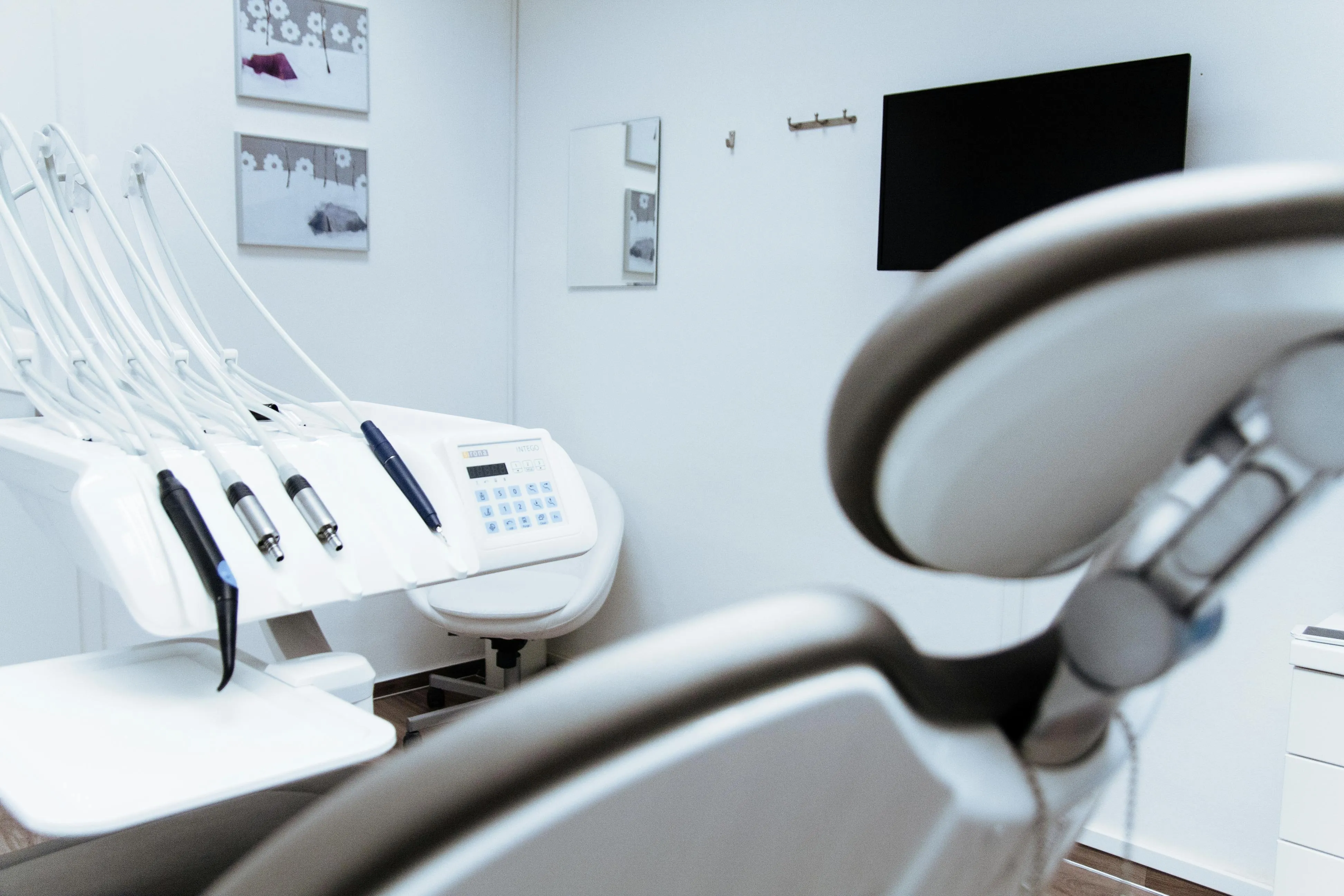
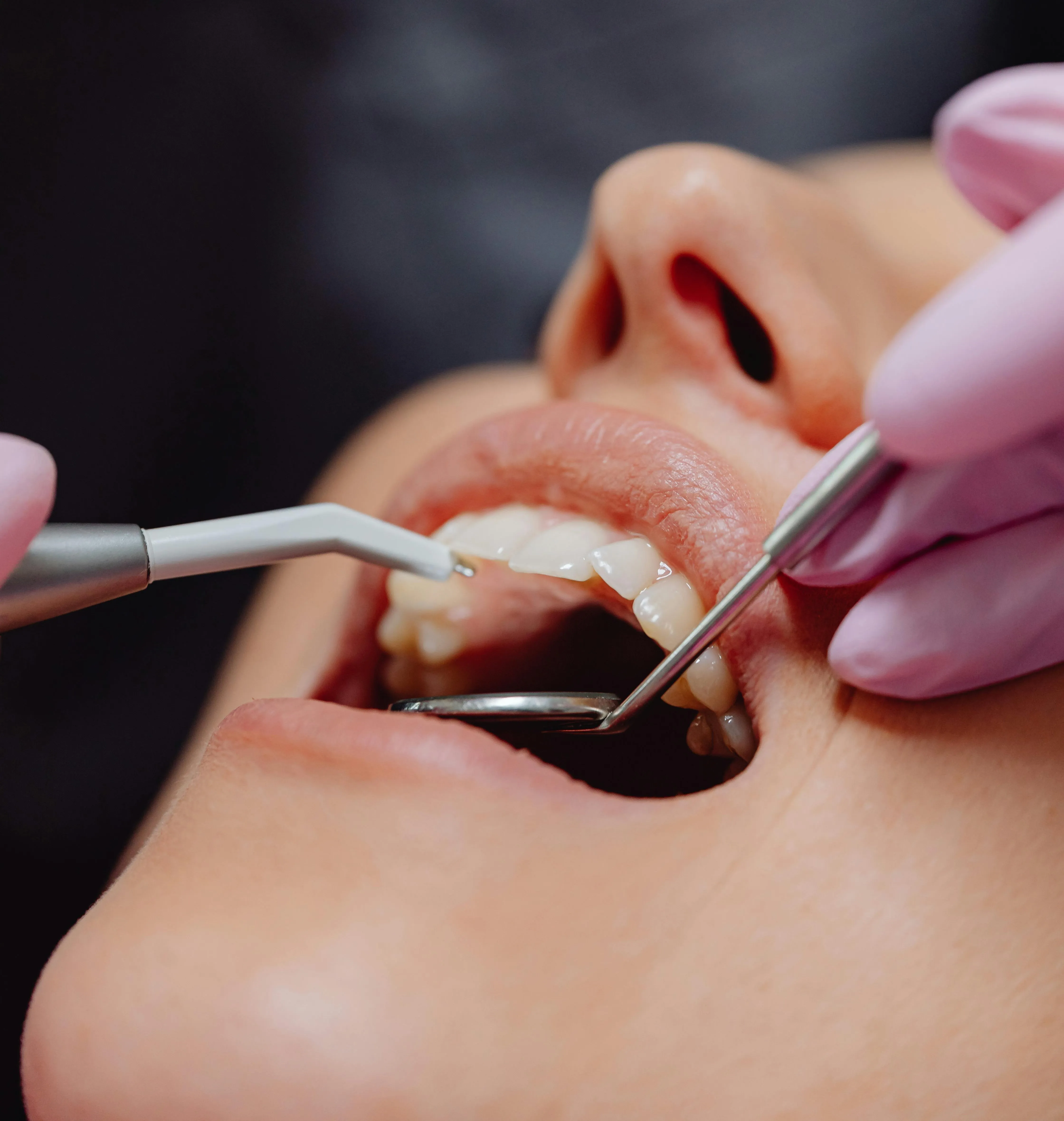
Illustrative images — neither belongs to a patient of this practice, and they are not the same mouth. Radiograph: Ruhrfisch, Wikimedia Commons, CC BY-SA 4.0. Photograph: Faisal Ghaffar, Pexels.
Treatments
How each treatment works, the conditions under which it is used and the cases in which it is not are explained on its own page.
Dental ImplantA titanium screw placed in the jawbone to replace a lost tooth, with a crown built on top of it.
OrthodonticsCorrecting crowded teeth or a faulty bite with braces or clear aligners.
Teeth WhiteningLightening the tooth's own colour by a few shades without harming its structure.
Root Canal TreatmentCleaning out the inflamed nerve tissue and filling the canal — saving the tooth instead of extracting it.
Zirconia CrownA crown with no metal substructure, whose translucency is close to that of a natural tooth.
Paediatric DentistryTreating baby teeth, preventing decay, and building the child's relationship with the dentist.
During treatment
The dentist who carries out the treatment
Dt. Ahmet Alper Güler. From the examination through to the completion of treatment, the process is carried out by one dentist.
The practice